The small intestine is the principal site where the bulk of chemical digestion is completed and nutrients are transferred into the body’s internal milieu. Although it functions as a continuous tube, it is traditionally divided into three distinct anatomical and functional regions: the duodenum, the jejunum, and the ileum. Each segment possesses unique structural features, enzymatic environments, and absorptive specializations that together ensure efficient breakdown of macronutrients, micronutrient uptake, and preparation of luminal contents for entry into the portal circulation and lymphatic system. Understanding the nuances of these three sections is essential for anyone studying digestive physiology, nutrition science, or clinical gastroenterology.
Duodenum: The Chemical Hub of Digestion
Anatomical Layout
The duodenum is the shortest portion of the small intestine, extending roughly 25–30 cm from the pyloric sphincter of the stomach to the ligament of Treitz, where it transitions into the jejunum. It forms a C‑shaped curve that lies retroperitoneally, allowing close proximity to the pancreas, liver, and gallbladder. This anatomical relationship is not incidental; it underpins the duodenum’s role as the primary site for the mixing of gastric chyme with pancreatic juice, bile, and intestinal secretions.
Mucosal Architecture
While the duodenal mucosa shares the typical villus‑crypt organization of the small intestine, it exhibits a higher density of Brunner’s glands within the submucosa. These glands secrete an alkaline mucus rich in bicarbonate ions, which neutralizes the acidic chyme arriving from the stomach (pH ≈ 2–3) and raises the luminal pH to around 6–7. This pH shift is critical for optimal activity of pancreatic enzymes and for protecting the epithelial surface from acid injury.
Enzymatic and Secretory Interplay
The duodenum is the principal arena for several key digestive processes:
- Pancreatic Enzyme Activation: Trypsinogen, chymotrypsinogen, procarboxypeptidases, and proelastase are secreted by the pancreas in inactive forms. Enterokinase (also called enteropeptidase), a brush‑border enzyme expressed on duodenal enterocytes, cleaves trypsinogen to active trypsin, which then autocatalytically activates the remaining zymogens. This cascade initiates protein hydrolysis.
- Bile Salt Emulsification: Bile, stored in the gallbladder and released via the common bile duct, contains bile salts that emulsify dietary lipids into micelles. The duodenum’s turbulent flow, enhanced by peristaltic mixing, maximizes the surface area of lipid droplets, facilitating lipase access.
- Brush‑Border Enzyme Activity: Disaccharidases (e.g., lactase, sucrase‑isomaltase) and peptidases are densely expressed on the duodenal microvilli, beginning the final steps of carbohydrate and peptide digestion before the chyme proceeds downstream.
Regulatory Hormones
Two duodenal hormones dominate early post‑prandial regulation:
- Secretin: Released in response to low pH, secretin stimulates pancreatic ductal cells to secrete bicarbonate‑rich fluid, reinforcing luminal alkalinization.
- Cholecystokinin (CCK): Triggered by the presence of fatty acids and amino acids, CCK prompts gallbladder contraction (releasing bile) and pancreatic acinar secretion (releasing digestive enzymes). It also slows gastric emptying, allowing the duodenum sufficient time to process incoming chyme.
Jejunum: The Primary Site of Nutrient Absorption
Anatomical Characteristics
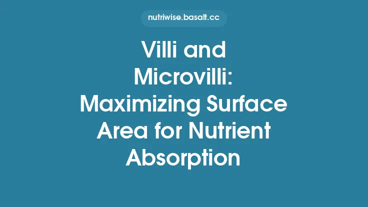
The jejunum comprises roughly the next two‑thirds of the small intestine, extending from the ligament of Treitz to the distal ileum. It is typically located in the left upper quadrant of the abdominal cavity and is distinguished by its thicker wall, broader lumen, and more prominent plicae circulares (circular folds) compared to the ileum. These folds increase the mucosal surface area by up to 10‑fold, optimizing absorptive capacity.
Mucosal Specializations
The jejunal mucosa exhibits:
- Longer Villi and Deeper Crypts: While the villus‑crypt pattern is universal, jejunal villi are generally taller, and crypts are deeper, supporting a higher turnover rate of enterocytes and a larger absorptive surface.
- Enhanced Brush‑Border Enzyme Density: Enzymes such as maltase, isomaltase, and sucrase are highly expressed, facilitating rapid disaccharide hydrolysis. Peptidases (aminopeptidases, dipeptidases) are also abundant, completing protein digestion.
Absorptive Mechanisms
The jejunum is the principal location for the uptake of most macronutrients and many micronutrients:
- Carbohydrates: Monosaccharides (glucose, galactose, fructose) are absorbed via specific transporters. Sodium‑glucose linked transporter 1 (SGLT1) mediates active uptake of glucose and galactose coupled to Na⁺, while facilitated diffusion via GLUT2 handles fructose and the efflux of glucose into the portal blood.
- Amino Acids and Small Peptides: Multiple Na⁺‑dependent amino acid transporters (e.g., B⁰AT1, PAT1) and peptide transporters (PEPT1) enable efficient uptake of amino acids and di‑/tripeptides.
- Lipids: Long‑chain fatty acids and monoglycerides, after micellar solubilization, diffuse across the enterocyte membrane, re‑esterify into triglycerides, and are packaged into chylomicrons for lymphatic transport via lacteals.
- Vitamins and Minerals: Water‑soluble vitamins (e.g., B‑complex, C) are absorbed by carrier‑mediated processes, while certain fat‑soluble vitamins (A, D, E, K) follow the lipid absorption pathway. Iron (Fe²⁺) uptake is facilitated by divalent metal transporter 1 (DMT1) in the jejunal epithelium.
Motility Patterns
Jejunal peristalsis is characterized by strong, coordinated contractions that propel chyme while segmentation contractions mix contents, ensuring intimate contact with the absorptive surface. The intrinsic smooth‑muscle activity is modulated by the myenteric plexus, but the focus here is on the mechanical outcome: efficient mixing and transit.
Ileum: The Final Absorptive Frontier and Immune Sentinel
Anatomical Positioning
The ileum occupies the distal third of the small intestine, extending from the jejuno‑ileal junction to the ileocecal valve. It is generally longer (≈ 3–4 m) but has a narrower lumen and thinner wall than the jejunum. Its location in the right lower quadrant brings it into close proximity with the cecum and colon.
Distinct Morphology
Key features that differentiate the ileum include:
- Peyer’s Patches: Aggregates of lymphoid tissue embedded in the submucosa, representing the gut‑associated lymphoid tissue (GALT). These structures monitor luminal antigens and facilitate the generation of IgA‑producing plasma cells, contributing to mucosal immunity.
- Fewer and Shorter Villi: The ileal villi are comparatively shorter, reflecting a shift from maximal absorptive surface to specialized functions.
- Higher Density of Specific Transporters: The ileum expresses transporters for nutrients that are less efficiently absorbed earlier, such as bile acids and vitamin B₁₂.
Specialized Absorptive Functions
- Bile Acid Reabsorption: The ileum is the primary site for enterohepatic recirculation of bile acids. The apical sodium‑dependent bile acid transporter (ASBT) reclaims conjugated bile acids from the lumen, returning them to the portal circulation via the ileal bile acid binding protein (IBABP) and the organic solute transporter α/β (OSTα/β). Approximately 95 % of bile acids are reclaimed, conserving the bile acid pool.
- Vitamin B₁₂–Intrinsic Factor Complex: Vitamin B₁₂, after binding intrinsic factor (IF) in the stomach, is absorbed in the distal ileum via the cubilin–amnionless receptor complex. This highly specific uptake mechanism is essential for hematopoiesis and neurologic function.
- Residual Nutrient Absorption: Any remaining monosaccharides, amino acids, and short‑chain fatty acids (SCFAs) that escaped earlier absorption are taken up here, albeit at lower rates.
Regulatory Hormones and Paracrine Signals
The ileum secretes several hormones that influence gastrointestinal function:
- Glucagon‑Like Peptide‑1 (GLP‑1) and Glucose‑Dependent Insulinotropic Polypeptide (GIP): Both are incretin hormones released by enteroendocrine L‑cells (GLP‑1) and K‑cells (GIP) in response to nutrient exposure. They potentiate insulin secretion, modulate gastric emptying, and affect appetite regulation.
- Peptide YY (PYY): Also released by L‑cells, PYY slows intestinal motility and contributes to satiety signaling.
These hormones illustrate how the ileum participates in systemic metabolic homeostasis beyond its local absorptive duties.
Comparative Anatomy and Functional Specialization
| Feature | Duodenum | Jejunum | Ileum |
|---|---|---|---|
| Length (approx.) | 25–30 cm | 2–3 m | 3–4 m |
| Primary Role | Chemical digestion, pH neutralization, enzyme activation | Bulk nutrient absorption (carbohydrates, amino acids, lipids) | Bile acid & vitamin B₁₂ absorption, immune surveillance |
| Mucosal Structures | Brunner’s glands, high bicarbonate secretion | Prominent plicae circulares, tall villi | Peyer’s patches, shorter villi |
| Key Transporters | Enterokinase, SGLT1 (early glucose uptake) | SGLT1, GLUT2, PEPT1, various amino acid transporters | ASBT (bile acids), cubilin‑IF complex (B₁₂), GLP‑1/GIP secretion |
| Hormonal Influence | Secretin, CCK (stimulate pancreatic/biliary secretions) | Minimal direct hormonal output | GLP‑1, GIP, PYY (systemic metabolic effects) |
| Motility Pattern | Strong peristalsis to move chyme rapidly | Segmentation and peristalsis for mixing/absorption | Slower peristalsis, allowing maximal extraction of residual nutrients |
The gradient of function—from aggressive chemical processing in the duodenum to refined absorptive and immunologic tasks in the ileum—reflects evolutionary optimization for nutrient extraction while preserving homeostatic balance.
Hormonal and Enzymatic Coordination Across Segments
The seamless transition from one intestinal segment to the next is orchestrated by a tightly regulated network of hormones, enzymes, and feedback loops:
- Acidic Chyme Arrival → Secretin Release → Pancreatic Bicarbonate Secretion → Duodenal pH Normalization.
- Fatty Acids & Amino Acids in Duodenum → CCK Release → Gallbladder Contraction + Pancreatic Enzyme Release → Enhanced Lipid Emulsification & Proteolysis.
- Glucose & Lipid Sensing in Jejunum → Incretin Release (GLP‑1, GIP) → Pancreatic β‑cell Stimulation → Insulin Secretion → Peripheral Glucose Uptake.
- Bile Acid Reabsorption in Ileum → FXR (Farnesoid X Receptor) Activation → Negative Feedback to Hepatic Bile Acid Synthesis → Maintenance of Bile Acid Pool.
These intersegmental signals ensure that digestive secretions are matched to luminal demands, preventing wasteful over‑production and protecting the mucosa from excessive acidity or enzymatic activity.
Clinical Considerations Linked to Segmental Dysfunction
While the article’s focus is anatomical and physiological, awareness of segment‑specific pathologies underscores the importance of each region:
- Duodenal Ulceration: Persistent exposure to gastric acid can breach the protective bicarbonate layer, leading to ulcer formation. Understanding duodenal secretory mechanisms informs therapeutic use of proton‑pump inhibitors and H₂‑blockers.
- Jejunal Malabsorption: Conditions such as celiac disease or tropical sprue primarily affect the jejunum, resulting in steatorrhea, weight loss, and deficiencies in iron, folate, and fat‑soluble vitamins. Histologic assessment often reveals villous atrophy specific to this segment.
- Ileal Resection: Surgical removal of the terminal ileum (e.g., for Crohn’s disease) impairs bile acid reabsorption, causing bile acid diarrhea, and disrupts vitamin B₁₂ uptake, leading to megaloblastic anemia. Post‑resection management includes bile acid sequestrants and B₁₂ supplementation.
- Peyer’s Patch Hyperplasia: Overactivation of GALT can be seen in certain infections or immunologic disorders, potentially contributing to chronic inflammation and altered motility.
These examples illustrate how the distinct functions of each segment translate into specific clinical manifestations when disrupted.
Emerging Research and Future Directions
Recent advances are expanding our understanding of small‑intestinal segmental biology:
- Organoid Models: Human duodenal, jejunal, and ileal organoids derived from stem cells now permit segment‑specific investigation of nutrient transport, drug metabolism, and host‑microbe interactions in vitro.
- Microbiome Gradients: While the colon harbors the densest microbial community, the small intestine exhibits a subtle but meaningful gradient of bacterial populations, with the ileum hosting higher densities than the duodenum. Ongoing studies explore how these microbes influence bile acid metabolism and immune education via Peyer’s patches.
- Targeted Drug Delivery: Enteric coating technologies are being refined to release therapeutics at precise intestinal locations (e.g., duodenal‑targeted GLP‑1 analogs, ileal‑released bile acid sequestrants), leveraging segment‑specific pH and transit times.
- Nutrigenomics: Genetic variations in transporters such as SGLT1, PEPT1, and ASBT are being linked to individual differences in nutrient absorption efficiency, opening avenues for personalized nutrition strategies.
These frontiers promise to deepen our grasp of how each intestinal segment contributes to overall health and disease.
Summary
The small intestine’s three segments—duodenum, jejunum, and ileum—form a coordinated cascade of digestive and absorptive processes:
- The duodenum acts as the chemical hub, neutralizing gastric acid, activating pancreatic enzymes, and emulsifying fats through bile.
- The jejunum provides the bulk of nutrient absorption, equipped with extensive villi, high enzyme density, and specialized transporters for carbohydrates, amino acids, and lipids.
- The ileum finalizes absorption, reclaiming bile acids, capturing vitamin B₁₂, and serving as an immunologic sentinel via Peyer’s patches, while also secreting hormones that influence systemic metabolism.
Understanding the distinct anatomy, physiology, and regulatory mechanisms of each segment not only enriches foundational knowledge in food science and digestion but also informs clinical practice, nutritional counseling, and emerging biomedical research.